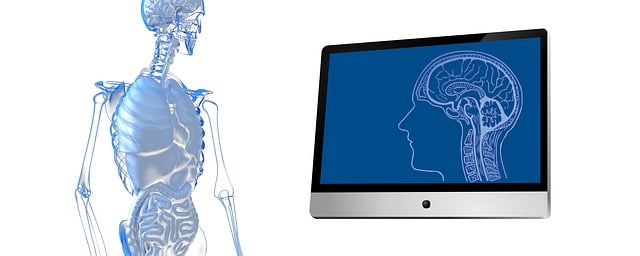
Advanced brain imaging technologies, especially magnetic resonance imaging (MRI), are crucial for detecting multiple sclerosis (MS) through visualization of cerebral cortex changes. Early detection via stroke diagnosis imaging enables timely interventions to slow MS progression and preserve neurological health by accurately identifying lesions and structural modifications.
Brain imaging technologies have revolutionized the way multiple sclerosis (MS) is detected and managed. By unveiling subtle changes within the brain, advanced stroke diagnosis imaging techniques play a pivotal role in identifying MS lesions. This article explores how these cutting-edge tools, including magnetic resonance imaging (MRI) and diffusion tensor imaging (DTI), facilitate early detection, track disease progression, and personalize treatment plans for MS patients.
Unveiling Brain Changes: Stroke Diagnosis Imaging Techniques
Brain imaging plays a pivotal role in unraveling subtle changes within the cerebral cortex, crucial for accurate multiple sclerosis (MS) detection. Techniques such as magnetic resonance imaging (MRI) offer a glimpse into the brain’s structure and function, enabling healthcare professionals to identify abnormalities indicative of MS. These changes may include demyelination, inflammation, and neurodegeneration, which are often difficult to discern through traditional means.
Stroke diagnosis imaging techniques, including computed tomography (CT) and MRI, provide dynamic visuals of cerebral blood flow and tissue integrity. By comparing these images with those from healthy individuals, researchers can pinpoint alterations specific to MS. This early detection is invaluable, as it allows for prompt intervention and management, potentially slowing the progression of the disease and preserving neurological function.
Detecting MS Lesions: Advanced Imaging Technologies
Brain imaging technologies have significantly advanced in recent years, revolutionizing the way we detect and diagnose multiple sclerosis (MS). Traditional methods often relied on symptoms and neurological examinations, but modern stroke diagnosis imaging offers a more precise approach. Techniques like magnetic resonance imaging (MRI) can uncover subtle changes in brain structure caused by MS lesions. These lesions are areas of damage or inflammation in the brain and spinal cord, which are characteristic of the disease.
Advanced MRI scanners, including those with high-resolution capabilities and specialized sequences, enable healthcare professionals to identify these lesions more effectively. By visualizing the brain’s intricate details, doctors can pinpoint the location, size, and number of lesions, aiding in an accurate MS diagnosis. This early detection is crucial as it allows for timely intervention and better management of the disease progression.
Enhancing Visualization: Tracking Disease Progression
Brain imaging plays a pivotal role in enhancing visualization and tracking disease progression in multiple sclerosis (MS). Advanced techniques like magnetic resonance imaging (MRI) allow healthcare professionals to peer into the brain, revealing subtle changes that may indicate MS activity. By comparing scans over time, doctors can monitor the extent of damage, track the progression of the disease, and even predict future relapses or disabilities with greater accuracy.
This capability is particularly crucial in differentiating MS from other conditions that share similar symptoms, such as stroke diagnosis imaging. The dynamic nature of MRI scans enables the detection of active lesions, demyelinization, and neurodegeneration—hallmarks of MS—which can help tailor treatment plans to address specific areas affected by the disease. This detailed visualization not only aids in early detection but also facilitates more precise interventions, ultimately improving patient outcomes.
Personalized Treatment: Brain Imaging's Role in MS Care
In the realm of multiple sclerosis (MS) care, brain imaging plays a pivotal role in personalized treatment strategies. By utilizing advanced imaging techniques such as magnetic resonance imaging (MRI), healthcare professionals can now peer into the intricate details of the brain, enabling them to make more accurate diagnoses and tailor treatments accordingly. This is particularly crucial in MS, where early detection and precise management are essential for mitigating symptoms and slowing disease progression.
Through stroke diagnosis imaging methods, brain scanning helps identify lesions and structural changes indicative of MS activity. These insights facilitate personalized care plans, ensuring that each patient receives the most effective therapies targeted at specific symptoms and disease modifications. This approach enhances treatment outcomes, improves quality of life, and offers hope for a more proactive management strategy in the complex landscape of MS.
Brain imaging technologies have revolutionized multiple sclerosis (MS) detection, offering valuable insights into brain changes and disease progression. Advanced imaging techniques, such as stroke diagnosis imaging methods, enable early identification of MS lesions, facilitating personalized treatment plans. By enhancing visualization, healthcare professionals can better understand the unique neurologic landscape of each patient, ultimately improving care outcomes for those affected by MS.
