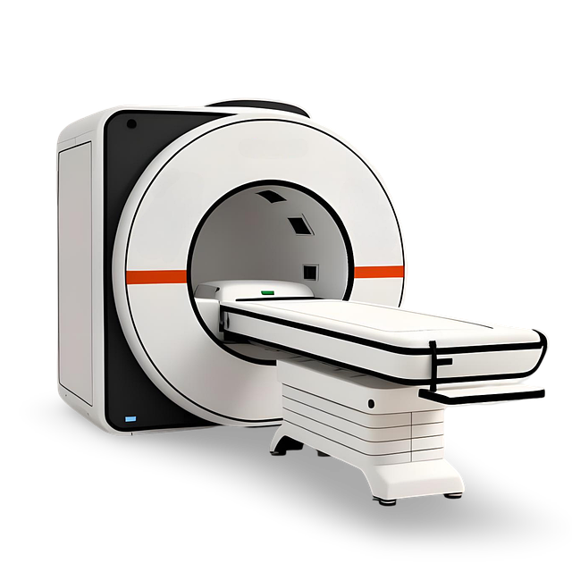
Electroencephalography (EEG) captures brain activity through scalp electrodes, offering real-time insights into cognitive processes and brain health but limited spatial resolution. Magnetic Resonance Imaging (MRI) provides detailed 3D structural views with superior spatial detail but lower temporal resolution and higher costs. Combining EEG with MRI for brain tumor imaging offers a comprehensive approach, leveraging the strengths of both techniques to aid diagnosis and treatment decisions by presenting both functional and structural information. Understanding the advantages and limitations of these methods is crucial for effective brain tumor management.
In the realm of brain health, understanding different imaging techniques is paramount. This article delves into the nuances of two prominent methods: Electroencephalography (EEG) and brain imaging technologies. While EEG captures neural activity through electrical signals, brain imaging employs various modalities to visualize structural and functional aspects of the brain. When it comes to detecting brain tumors, comparing these techniques reveals unique advantages and limitations. By exploring their roles and differences, especially in brain tumor imaging, readers gain insights into optimal diagnostic strategies.
Understanding Electroencephalography (EEG)
Electroencephalography (EEG) is a non-invasive technique that records electrical activity in the brain using electrodes placed on the scalp. This method captures the brain’s rhythmic patterns, known as brain waves, which vary according to different mental states and activities. By analyzing these waves, researchers and medical professionals can gain insights into brain function, diagnosis, and treatment monitoring. EEG is particularly valuable for studying cognitive processes, sleep disorders, epilepsy, and even brain tumor imaging, making it a powerful tool in neurology.
The data obtained from EEG provides information on neural communication, allowing scientists to identify abnormalities associated with various neurological conditions. In the context of brain tumor imaging, EEG can help detect irregular brain activity caused by tumors, providing critical information for diagnosis and treatment planning. This technology’s ability to offer real-time feedback makes it an essential component in understanding and managing brain health.
The Role of Brain Imaging Technologies
Brain imaging technologies play a pivotal role in the field of neuroscience and medical diagnosis. These advanced tools enable researchers and healthcare professionals to peer into the complex workings of the brain, offering invaluable insights into its structure, function, and any abnormalities that may arise. From detecting subtle changes indicative of neurological disorders to guiding treatment plans for conditions like brain tumors, brain imaging is an indispensable asset in modern medicine.
One such technology, magnetic resonance imaging (MRI), uses strong magnetic fields and radio waves to generate detailed images of the brain’s anatomy. This non-invasive technique allows doctors to visualize brain tumors, study their size, location, and impact on surrounding areas, thereby facilitating informed decisions for surgical interventions or radiation therapy. Other imaging methods, such as computed tomography (CT) scans, utilize X-rays to create cross-sectional images, providing rapid assessments of traumatic injuries, hemorrhages, or congenital anomalies, including those affecting the brain.
Comparing EEG and Brain Imaging for Brain Tumor Detection
When it comes to brain tumor imaging, both Electroencephalography (EEG) and conventional brain imaging techniques like magnetic resonance imaging (MRI) offer valuable insights into brain health. However, they differ significantly in their approaches. EEG measures the electrical activity of the brain through electrodes placed on the scalp, capturing dynamic changes in real-time. This non-invasive method is particularly useful for assessing functional abnormalities associated with tumors and their impact on neural networks.
In contrast, traditional brain imaging such as MRI provides detailed structural information by generating high-resolution images of the brain’s anatomy. While it excels at identifying the presence, size, and location of a tumor, EEG offers a more direct window into the electrical signaling that may be disrupted by tumors. Combining these two techniques can provide a comprehensive understanding of a patient’s condition, guiding treatment decisions for effective brain tumor management.
Advantages and Limitations of Each Method
Advantages and Limitations of Each Method
EEG (Electroencephalography)
One of the primary advantages of EEG is its non-invasiveness, making it a preferred method for many research and clinical applications. It can capture brain activity in real-time, providing insights into dynamic processes like cognitive functions and sleep patterns. Moreover, EEG offers high temporal resolution, allowing researchers to observe changes in brainwaves within milliseconds. However, EEG has limitations when it comes to spatial resolution. It measures the average electrical activity over a large number of neurons, making it challenging to pinpoint specific brain regions or structures, especially smaller ones like those affected by a brain tumor imaging.
Brain Imaging (e.g., MRI, fMRI)
Brain imaging techniques, such as Magnetic Resonance Imaging (MRI) and functional Magnetic Resonance Imaging (fMRI), offer superior spatial resolution compared to EEG. They can produce detailed, three-dimensional maps of the brain, revealing structural anomalies or changes in blood flow indicative of neurological conditions or diseases, including brain tumors. These methods are non-invasive and safe for repeated use. However, they have lower temporal resolution than EEG, as they require longer acquisition times and cannot capture real-time dynamic changes in brain activity. Additionally, MRI and fMRI can be more expensive and accessible only in specialized facilities.
In the quest for accurate brain tumor imaging, both Electroencephalography (EEG) and advanced brain imaging technologies offer unique insights. While EEG provides a non-invasive method to record brain activity, brain imaging technologies like MRI and PET scans excel in visualizing structural abnormalities. When comparing EEG and brain imaging for brain tumor detection, each method has its advantages and limitations. EEG is quick, cost-effective, and useful for monitoring brainwaves during various cognitive tasks, making it valuable for neurological assessments. On the other hand, brain imaging technologies offer high-resolution visualizations of the brain’s anatomy, helping in diagnosing and staging brain tumors. Understanding these differences is crucial for healthcare professionals to choose the most appropriate tool for effective brain tumor imaging and treatment planning.
