Electroencephalography (EEG) and Functional Magnetic Resonance Imaging (fMRI) are complementary brain imaging techniques for stroke diagnosis imaging. EEG detects rapid electrical changes in neural networks, while fMRI tracks blood flow to identify areas of reduced supply. EEG offers real-time monitoring, non-invasiveness, affordability, and ease of use, but has low spatial resolution. fMRI provides superior spatial resolution but is more expensive, requires longer patient immobility, and may have contraindications. Both methods are essential for swift stroke assessment and tailored treatment plans.
In the realm of brain imaging, Electroencephalography (EEG) and Functional Magnetic Resonance Imaging (fMRI) stand out as powerful tools for understanding cerebral function. While EEG captures brain waves through electrical signals, fMRI tracks blood flow changes, offering unique insights into neural activity. This article delves into the intricacies of these techniques, focusing on stroke diagnosis, their respective advantages and limitations, and how each method contributes to our understanding of the brain, with a special emphasis on stroke imaging applications.
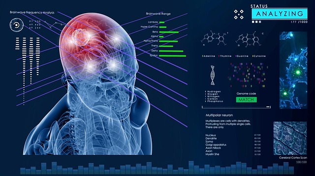
Electroencephalography (EEG): Capture Brain Waves
Electroencephalography (EEG) is a non-invasive brain imaging technique that records electrical activity in the brain through electrodes placed on the scalp. This method captures brain waves, or electroencephalograms, which are measured in Hertz (cycles per second). EEG is particularly useful for stroke diagnosis imaging as it can detect changes in brain function and electrical patterns associated with cerebral events. By analyzing these waves, healthcare professionals can gather insights into cognitive performance, brain damage, and recovery processes.
EEG offers high temporal resolution, allowing for the observation of dynamic brain activity in real-time. This makes it invaluable for monitoring brain responses to various stimuli, tracking sleep patterns, and studying seizures or epilepsy. Unlike some other imaging methods, EEG is relatively affordable and easily accessible, making it a preferred choice for many clinical and research applications, especially when quick assessment and continuous monitoring are required, such as in stroke diagnosis and care.
Functional Magnetic Resonance Imaging (fMRI): Blood Flow Tracking
Functional Magnetic Resonance Imaging (fMRI) is a powerful tool in brain imaging that tracks changes in blood flow, allowing researchers to monitor neural activity. Unlike EEG, which records electrical signals directly from the brain’s surface, fMRI indirectly measures brain activity by detecting the increase in blood flow to active areas. This method provides valuable insights into the brain’s functional connectivity and is widely used in stroke diagnosis imaging. By identifying areas of reduced or altered blood flow, healthcare professionals can pinpoint regions affected by a stroke, aiding in more accurate patient assessment and personalized treatment planning.
Stroke Diagnosis: EEG vs. fMRI Comparison
When it comes to stroke diagnosis, different brain imaging techniques offer unique insights. Electroencephalography (EEG) focuses on electrical activity in the brain, capturing rapid changes in neural networks. This makes EEG valuable for detecting acute changes associated with strokes, particularly in the early stages when structural damage might not be evident on other scans.
In contrast, functional Magnetic Resonance Imaging (fMRI) tracks blood flow to measure brain activity. While fMRI provides a broader view of cerebral function and can identify areas of the brain affected by reduced blood flow or ischemia, it’s less sensitive to the initial electrical disruptions that EEG picks up. This difference makes EEG a critical tool for rapid assessment and monitoring of stroke patients, helping healthcare providers make timely interventions.
Advantages and Limitations of Each Method
EEG vs Brain Imaging: Advantages and Limitations
Electroencephalography (EEG) offers a unique perspective in stroke diagnosis imaging. Its primary advantage lies in capturing real-time brain activity, making it excellent for monitoring neurological conditions and detecting subtle changes that may occur immediately after a stroke. EEG is non-invasive, affordable, and relatively easy to perform, allowing for frequent assessments without causing discomfort to the patient. However, its limitations include low spatial resolution, making it challenging to pinpoint precise brain regions affected by a stroke. Moreover, EEG recordings can be influenced by muscle artifacts and movements, which may obscure the interpretation of signals from the brain.
On the other hand, brain imaging techniques like magnetic resonance imaging (MRI) provide superior spatial resolution, enabling healthcare professionals to visualize structural changes within the brain with greater detail. Stroke diagnosis using MRI is invaluable for identifying infarct size, location, and potential vascular complications. However, MRI scanners are more expensive and require patients to remain still for extended periods. Additionally, some individuals may have contraindications due to metal implants or other medical conditions, limiting its accessibility in certain cases.
In comparing EEG and fMRI for stroke diagnosis, each method offers unique insights into brain function. Electroencephalography (EEG), by measuring brain waves, provides real-time information about neural activity, while functional Magnetic Resonance Imaging (fMRI) tracks blood flow changes to identify active brain regions. While EEG is non-invasive, portable, and suitable for continuous monitoring, fMRI offers higher spatial resolution and can detect subtle differences in brain function. Together, these techniques complement each other, enhancing our ability to diagnose and understand stroke impacts, ultimately improving patient care. SEO keywords: stroke diagnosis imaging.