Functional neuroimaging techniques like MRI and PET scans revolutionize both stroke diagnosis imaging and psychiatric research. By mapping brain activity and connectivity, these tools identify subtle changes caused by strokes or mental health disorders. Advanced methods such as fNIRS and EEG offer detailed visualization of neural networks, enabling personalized treatments for improved patient outcomes, especially in individuals with comorbid psychiatric conditions post-stroke.
Functional neuroimaging has emerged as a powerful tool in understanding and diagnosing psychiatric disorders. This innovative technology allows researchers to map brain activity, providing insights into the neural mechanisms underlying mental health conditions. In this article, we explore key aspects of functional neuroimaging for psychiatric disorders, focusing on stroke diagnosis imaging as a pivotal technique. We delve into advanced brain mapping methods and discuss how interpreting data from these scans can unlock crucial insights, paving the way for more effective treatments.
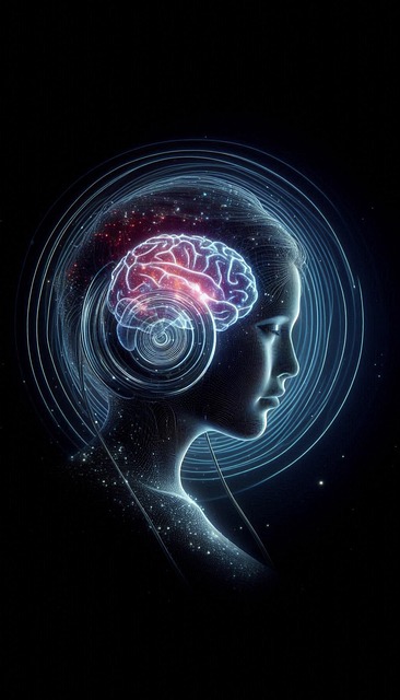
Understanding Functional Neuroimaging for Psychiatric Disorders
Functional neuroimaging offers a powerful window into understanding psychiatric disorders by mapping brain activity and connectivity. This non-invasive technique allows researchers to visualise which areas of the brain are activated during specific tasks or rest, providing insights into both typical and atypical brain function. By comparing patterns of brain activation between individuals with psychiatric conditions and healthy controls, scientists can identify distinctive neural signatures associated with various disorders.
Furthermore, functional neuroimaging has significant implications for stroke diagnosis imaging. It enables the detection of subtle changes in brain function resulting from stroke or other neurological events, even when structural damage may not be evident on traditional imaging scans. This early identification of dysfunctional brain regions can lead to more targeted interventions and improved patient outcomes.
Stroke Diagnosis Imaging: A Key Tool in Psychiatry
Stroke diagnosis imaging plays a pivotal role in psychiatric research, offering valuable insights into the complex neurobiology of mental health disorders. Functional neuroimaging techniques, such as magnetic resonance imaging (MRI) and positron emission tomography (PET), have revolutionized our understanding of brain function by allowing researchers to visualize and measure activity within living brains.
In psychiatry, these advanced imaging methods enable precise stroke diagnosis, tracking changes in brain structure and function over time, and identifying distinct neural circuits associated with various psychiatric conditions. By studying the brain’s response during cognitive tasks or emotional stimuli, researchers can uncover unique patterns that help differentiate between disorders, predict treatment outcomes, and ultimately, foster more personalized and effective therapeutic interventions for individuals struggling with mental health challenges.
Advanced Techniques in Brain Mapping for Mental Health
Advanced techniques in brain mapping have revolutionized functional neuroimaging, offering unprecedented insights into psychiatric disorders. These cutting-edge methods go beyond traditional stroke diagnosis imaging to uncover intricate neural networks and their dysfunctions. By employing tools such as magnetic resonance imaging (MRI), functional near-infrared spectroscopy (fNIRS), and electroencephalography (EEG), researchers can now visualize brain activity with remarkable detail, enabling a more nuanced understanding of mental health conditions.
For instance, fNIRS allows for non-invasive measurements of blood oxygen levels in the brain, correlating these signals with cognitive tasks to identify areas of heightened or diminished activity associated with specific psychiatric symptoms. Similarly, EEG provides real-time data on brain waves, aiding in the detection of abnormalities that may indicate disordered brain function. These advancements hold promise for personalized treatment approaches, as they can help clinicians pinpoint precise brain regions involved in a patient’s condition, paving the way for more targeted and effective interventions in the field of mental health.
Interpreting Data: Unlocking Insights into Psychiatric Disorders
Interpreting data from functional neuroimaging holds immense potential for unravelling the complexities of psychiatric disorders. By comparing patterns of brain activity between individuals with and without conditions like depression or schizophrenia, researchers can identify distinct neural circuits and networks associated with specific symptoms. This approach allows for a more nuanced understanding of each disorder’s underlying biology, moving beyond traditional diagnostic categories based solely on clinical presentation.
Moreover, advancements in stroke diagnosis imaging techniques enable the examination of brain structure and function after traumatic events. These studies contribute to our knowledge of recovery mechanisms and potential treatment targets, offering hope for improved outcomes in stroke survivors experiencing psychiatric comorbidities.
Functional neuroimaging has emerged as a powerful tool in understanding and diagnosing psychiatric disorders, offering insights that were previously unimaginable. From traditional methods like MRI to advanced techniques such as EEG and fMRI, these tools allow for precise brain mapping, enabling healthcare professionals to detect subtle differences associated with various mental health conditions. Specifically, stroke diagnosis imaging plays a pivotal role by providing critical information on brain function and structure, enhancing the accuracy of psychiatric diagnoses. As research continues to refine these techniques, the future looks promising for more effective treatment and improved outcomes in psychiatric care.