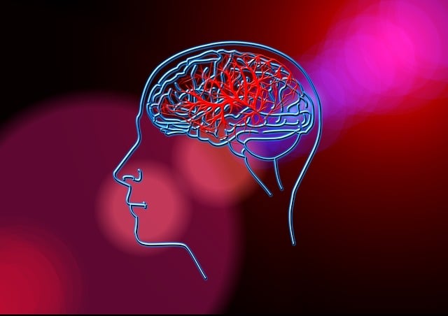
Brain CT scans are powerful diagnostic tools but carry radiation risks that accumulate over time. Brain PET scans offer insights into brain function with higher radiation doses. Radiation exposure during PET scans depends on tracer type and examined area. Multiple recurrent scans increase risk, requiring comprehensive assessment. Medical professionals minimize CT scan radiation through optimized parameters, protective gear, and patient education. Alternative techniques like PET scans and MRI reduce radiation concern, valuable for studying brain disorders.
In today’s medical landscape, brain PET scans offer crucial insights for diagnosis and treatment planning. However, concerns around radiation exposure are valid due to the sensitive nature of the brain. This article delves into understanding brain CT scans and their associated radiation risks, exploring factors impacting exposure during brain PET scans. We present safety measures to minimize these risks and discuss alternative imaging techniques that reduce radiation concern, providing a comprehensive guide for healthcare professionals and patients alike.
Understanding Brain CT Scans and Radiation Risk
Brain CT scans are a vital diagnostic tool, offering detailed images of the brain’s internal structures. However, like any medical imaging procedure, they carry a subtle risk of radiation exposure. It’s essential to grasp this concept, especially when considering repeated or alternative brain imaging methods like a brain PET scan.
The low-dose radiation used in CT scans accumulates over time, particularly in frequent or young patients. While the risks are generally low for a single scan, chronic exposure can lead to long-term health effects. Understanding these risks is crucial for informed consent and managing patient safety, especially when exploring advanced imaging techniques like brain PET scans which provide unique insights into brain function and metabolism but may expose individuals to higher radiation doses.
Factors Affecting Radiation Exposure During Brain PET Scans
Several factors influence the radiation exposure during a brain PET (Positron Emission Tomography) scan, and understanding them is essential for patients and healthcare providers alike. One significant factor is the type of tracer used in the scan. Different radioactive tracers have varying levels of radioactivity, which directly impacts the amount of radiation emitted during the procedure. Additionally, the specific brain area being examined plays a role; targeted scans focusing on particular regions may result in lower overall exposure compared to whole-brain assessments.
The duration and frequency of previous PET scans are also critical considerations. Subsequent scans, especially those performed within a short time frame, can accumulate radiation exposure over time. Patients with multiple recurrent scans might need more comprehensive risk assessment and potentially alternative imaging techniques to minimize radiation dose.
Minimizing Risks: Safety Measures for Brain CT Scans
Minimizing Risks: Safety Measures for Brain CT Scans
When it comes to brain CT scans, ensuring patient safety is paramount. Medical professionals employ various strategies to minimize radiation exposure during these critical examinations. One of the primary approaches involves optimizing scan parameters to deliver the lowest possible dose while still producing high-quality images. Modern scanners are equipped with advanced technology that allows for tailored settings based on the patient’s size and anatomy, thereby reducing unnecessary radiation.
Additionally, using lead shields and protective apparel can further decrease the amount of radiation absorbed by both the patient and medical staff. Patients are also educated about the importance of remaining still during the scan to prevent blurring or the need for repeat procedures, which would increase their overall radiation exposure. Regular maintenance and calibration of equipment ensure that these safety measures remain effective over time.
Alternative Imaging Techniques to Reduce Radiation Concern
In addition to optimizing scan parameters and minimizing exposure time, several alternative imaging techniques offer promising solutions to reduce radiation concern in brain CT scans. One such technique is the brain PET (Positron Emission Tomography) scan, which uses radioactive tracers to visualize metabolic processes in the brain. Unlike CT scans, PET scans have lower radiation doses because they capture functional information rather than anatomical details. This makes them a valuable alternative for diagnostic and research purposes, especially when studying brain disorders that involve changes in metabolism, like Alzheimer’s disease or Parkinson’s disease.
Moreover, magnetic resonance imaging (MRI) is another non-radiation based option that provides detailed images of the brain without exposure to ionizing radiation. MRI utilizes strong magnetic fields and radio waves to generate images, making it ideal for examining soft tissues and structural abnormalities. While MRI scans may take longer than CT scans, ongoing technological advancements continue to enhance their speed and resolution, further expanding their utility in neuroscientific research and clinical practice.
Brain CT scans, while crucial for diagnosing conditions affecting the brain, carry a risk of radiation exposure. By understanding the factors influencing this exposure and implementing safety measures, healthcare providers can minimize risks. Additionally, exploring alternative imaging techniques like brain PET scans offers further options to reduce radiation concern. Balancing diagnostic needs with patient safety is paramount in modern medicine, ensuring patients receive the best care while mitigating potential long-term effects of radiation exposure.
