Advanced medical imaging techniques like MRI, CT, and PET scans are vital for understanding Alzheimer's disease. These technologies reveal structural changes, metabolic shifts, and key hallmarks, aiding in early detection, accurate diagnosis, progression tracking, and assessment of treatment efficacy. Non-invasive brain imaging methods provide critical insights into cognitive decline and the evolution of AD over time, enabling healthcare professionals to tailor interventions and improve patient outcomes.
Medical imaging plays a pivotal role in diagnosing Alzheimer’s disease, revealing crucial brain changes associated with the degenerative condition. Through advanced techniques like MRI and PET scans, healthcare professionals can detect amyloid plugs and neurofibrillary tangles—hallmarks of the illness. These tools also enable measurement of brain atrophy, cerebral blood flow alterations, and tracking disease progression over time. By harnessing the power of medical imaging for brain assessment, doctors gain valuable insights into Alzheimer’s pathophysiology, facilitating earlier detection and more effective treatment strategies.
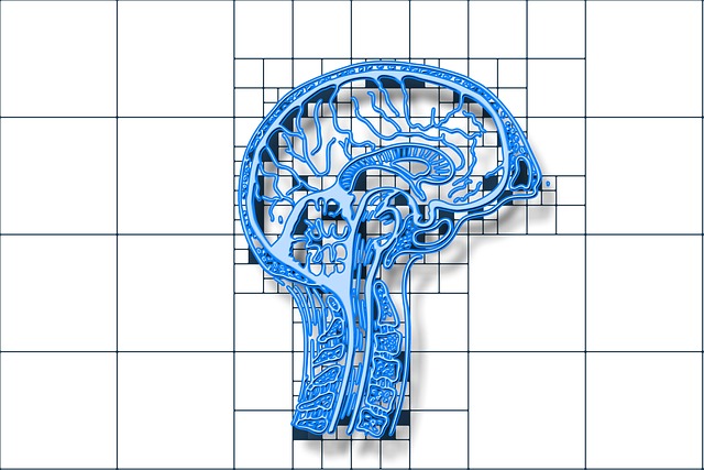
Unveiling Brain Changes: Medical Imaging Techniques
Medical imaging plays a pivotal role in unraveling the complex mysteries of Alzheimer’s disease by shedding light on subtle changes within the brain that might otherwise go unnoticed. Through advanced techniques like magnetic resonance imaging (MRI), computed tomography (CT), and positron emission tomography (PET), healthcare professionals can peer into the brain’s intricate architecture, identifying key indicators of the disease.
These imaging technologies enable visualization of structural abnormalities, such as nerve cell loss and shrinking brain volume, which are hallmarks of Alzheimer’s. Moreover, they facilitate detection of metabolic alterations and the buildup of amyloid plaques and tau proteins – toxic clumps that disrupt normal brain function and are strongly associated with the progression of the disease. By capturing these brain changes in high detail, medical imaging for brain becomes a powerful tool not only for diagnosis but also for tracking the disease’s course and evaluating the effectiveness of treatment interventions.
Detecting Amyloid Plugs and Neurofibrillary Tangles
Medical imaging plays a pivotal role in detecting key features of Alzheimer’s disease, such as amyloid plugs and neurofibrillary tangles. Advanced techniques like positron emission tomography (PET) and magnetic resonance imaging (MRI) enable healthcare professionals to visualize these structural abnormalities within the brain. PET scans, for instance, can pinpoint the accumulation of amyloid proteins, which are considered a hallmark of Alzheimer’s. Similarly, MRI technology helps identify neurofibrillary tangles—abnormal aggregates of tau protein that disrupt normal brain function.
By leveraging medical imaging for brain analysis, doctors can gain invaluable insights into the progression and severity of Alzheimer’s disease at various stages. This early detection not only aids in accurate diagnosis but also opens up opportunities for timely intervention and management strategies aimed at slowing down or even halting cognitive decline.
Measuring Atrophy and Cerebral Blood Flow
Medical imaging plays a pivotal role in diagnosing Alzheimer’s disease (AD) by providing detailed insights into the brain’s structure and function. One crucial aspect is the measurement of atrophy, or the loss of brain volume, which is a hallmark of AD. Advanced techniques like magnetic resonance imaging (MRI) allow healthcare professionals to track regional changes in brain size and identify areas of significant atrophy, helping to assess the progression of the disease.
Additionally, medical imaging aids in evaluating cerebral blood flow (CBF). Reduced CBF in specific regions has been linked to cognitive decline and AD symptoms. Functional MRI (fMRI) and computed tomography (CT) scans can detect changes in blood oxygenation and perfusion, providing valuable information about brain metabolic activity and potential impairments in blood vessel function associated with Alzheimer’s disease.
Tracking Disease Progression Over Time
Medical imaging plays a pivotal role in tracking the progression of Alzheimer’s disease over time. Through advanced techniques such as magnetic resonance imaging (MRI) and positron emission tomography (PET), healthcare professionals can non-invasively examine structural changes in the brain, identify key markers of the disease, and monitor its evolution. Regular imaging allows for early detection of cognitive decline, subtle atrophies in brain regions affected by Alzheimer’s, and even the buildup of amyloid plaques and tau proteins, which are hallmarks of the condition.
Over time, these images can show how the brain is changing, helping doctors assess the effectiveness of treatments and make informed decisions about patient care. This continuous monitoring is crucial for understanding disease progression, tailoring interventions to individual needs, and ultimately improving outcomes for those affected by Alzheimer’s. By leveraging medical imaging for brain analysis, researchers and clinicians gain valuable insights into the complex nature of this devastating disease.
Medical imaging plays a pivotal role in diagnosing Alzheimer’s disease, revealing key brain changes that were once invisible. Through advanced techniques, we can now detect amyloid plugs and neurofibrillary tangles, measure atrophy and cerebral blood flow, and track the progression of the disease over time. By harnessing the power of medical imaging for brain assessment, healthcare professionals gain invaluable insights to improve diagnosis and ultimately enhance patient care.