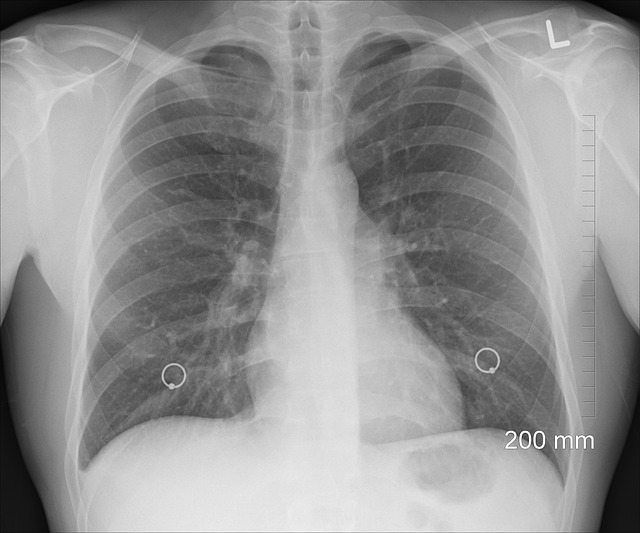
MRI offers detailed non-invasive insights into brain structure and function, crucial for neurologists. CT scans provide swift, high-resolution 3D models of cerebral vasculature and parenchyma, ideal for rapid diagnosis in emergencies. PET scans analyze brain function by tracking radiotracer compounds, aiding in understanding neurological disorders early on. Cerebral angiography visualizes blood flow within the brain's vessels using contrast agents, essential for diagnosing conditions like stroke, aneurysms, and vascular malformations.
Medical imaging plays a pivotal role in brain diagnosis, offering windows into its complex structure and function. From non-invasive techniques like Magnetic Resonance Imaging (MRI) that unveil brain anatomy, to functional analysis through Positron Emission Tomography (PET), each method provides unique insights. Computed Tomography (CT) scans deliver rapid assessments, while Cerebral Angiography visually maps blood flow, making it a crucial tool for understanding cerebral circulation. This comprehensive guide explores these diverse imaging types.
Magnetic Resonance Imaging (MRI): Unveiling Brain Structure
Magnetic Resonance Imaging (MRI) is a powerful tool in brain diagnosis, offering detailed insights into the structure and function of the cerebral tissue. This non-invasive technique employs strong magnetic fields and radio waves to generate precise images of the brain, allowing healthcare professionals to identify abnormalities that may indicate various neurological conditions. By providing high-resolution visuals of cerebral vasculature, MRI can reveal significant details about blood flow dynamics within the brain, a crucial aspect in diagnosing cerebrovascular diseases.
Unlike cerebral angiography, which involves injecting contrast agents to directly visualize blood vessels, MRI offers a more comprehensive view of the brain’s internal architecture. It can detect subtle changes in brain tissue structure, such as lesions, tumours, or areas of inflammation, making it invaluable for neurologists and neurosurgeons in their quest to pinpoint the source of cognitive or behavioural changes in patients.
Computed Tomography (CT) Scans: Rapid Assessment
Computed Tomography (CT) scans are a rapid and versatile tool for brain diagnosis, offering high-resolution cross-sectional images of the cerebral vasculature and parenchyma. This non-invasive procedure involves a series of X-rays taken from multiple angles to create detailed 3D models of the brain. CT scans can quickly identify bleeding, tumors, or inflammation in the brain, making them an essential first step in many diagnostic processes.
One specialized application is cerebral angiography, which uses contrast agents to visualize blood flow within the brain’s intricate network of vessels. This technique allows doctors to assess vascular abnormalities and make precise diagnoses, guiding treatment plans for conditions affecting the brain’s vasculature. CT scans’ speed and accuracy make them a go-to method for rapid assessment and initial evaluation in neurological emergencies.
Positron Emission Tomography (PET) for Functional Analysis
Positron Emission Tomography (PET) is a powerful tool for functional analysis of the brain, offering insights into its metabolic activity and blood flow patterns. This non-invasive imaging technique traces radiotracer compounds as they circulate through the body, allowing doctors to visualize and measure various physiological processes. In the context of cerebral angiography, PET scans can help identify blockages or abnormalities in blood vessels supplying the brain, providing crucial information for diagnosis and treatment planning.
By measuring glucose metabolism, PET scans can highlight areas of increased or decreased activity within the brain, offering a functional map that aids in understanding neurological disorders. This technology is particularly valuable in assessing conditions like Alzheimer’s disease, where it can detect changes in brain metabolism early on. Moreover, combining PET with other imaging modalities, such as magnetic resonance imaging (MRI), enhances diagnostic accuracy and offers a comprehensive view of both structural and functional aspects of the brain.
Cerebral Angiography: Visualizing Blood Flow in the Brain
Cerebral angiography is a specialized medical imaging technique that allows healthcare professionals to visualize blood flow within the intricate network of blood vessels in the brain. By injecting a contrast agent into an artery, typically in the neck or groin, this procedure offers a detailed look at the cerebral vasculature. The contrast agent highlights the blood vessels, making them easier to see on X-ray images or angiograms. This non-invasive method is crucial for diagnosing various conditions affecting blood flow to and within the brain, such as stroke, aneurysms, vascular malformations, and arteriovenous malformations (AVMs).
During cerebral angiography, a catheter is carefully maneuvered through the arteries to reach specific areas of interest in the brain. The contrast agent’s presence enhances the visibility of these structures, enabling doctors to assess their anatomy, identify blockages or leaks, and plan treatment strategies accordingly. This advanced imaging technique plays a pivotal role in providing accurate diagnoses and guiding interventions for conditions that impact the brain’s vascular system.
In conclusion, these diverse medical imaging techniques—Magnetic Resonance Imaging (MRI), Computed Tomography (CT) scans, Positron Emission Tomography (PET), and Cerebral Angiography—offer invaluable tools for brain diagnosis. Each method provides unique insights into different aspects of brain health, enabling healthcare professionals to make accurate assessments and develop tailored treatment plans. Understanding the strengths and applications of these technologies is essential in advancing neuroscience and improving patient care.
